Tistoryview
Disease&Treatment/Neuro-ophthalmology
visual pathway:optic tract, lateral geniculate body (LGB), optic radiations, and visual cortex.
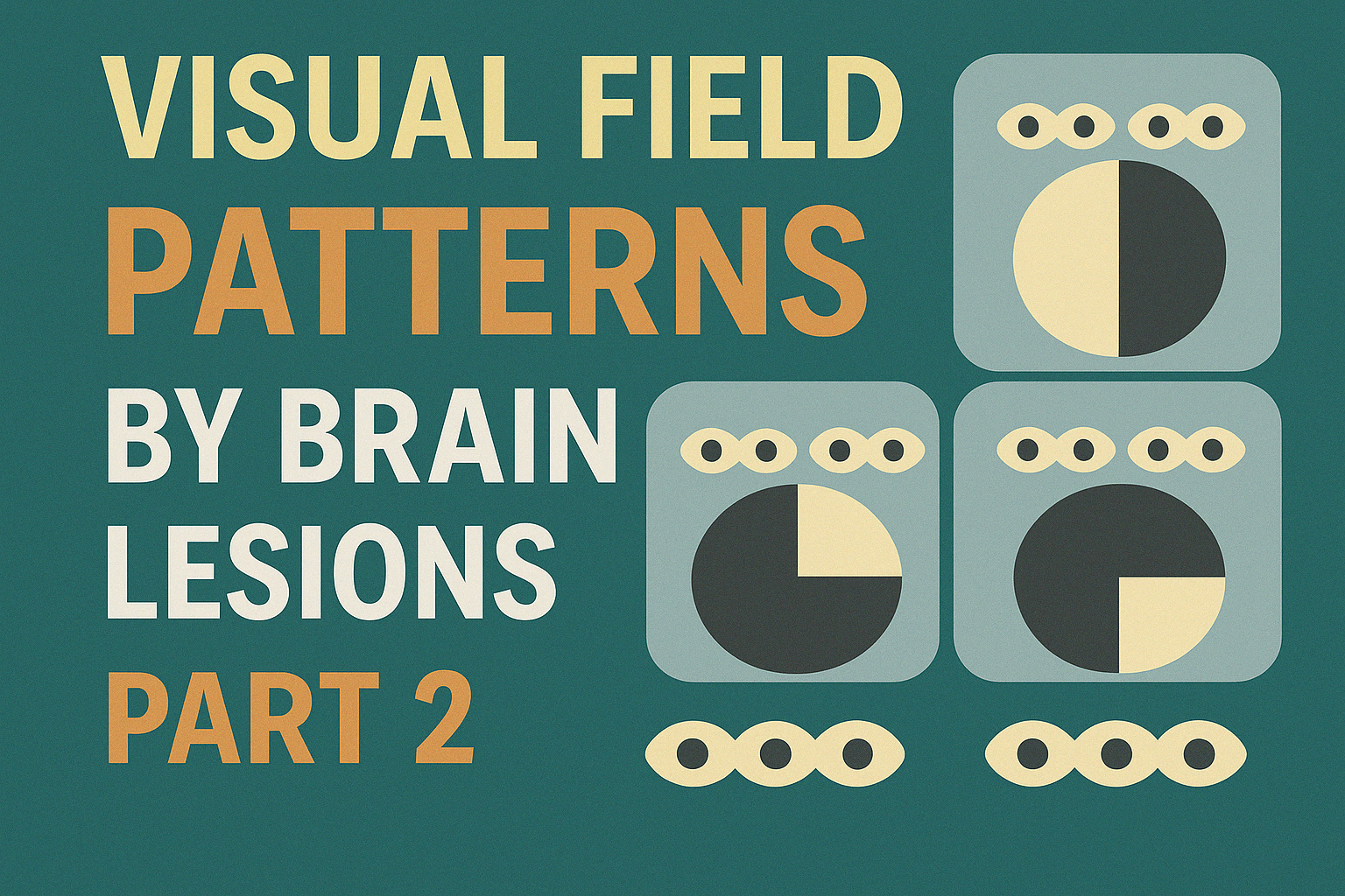
eye_doc 2025. 4. 19. 23:55👁 Visual Field Defects by Lesion Location (Part 2)
Following the previous overview of optic chiasm lesions,
this summary focuses on the next segments of the visual pathway:
optic tract, lateral geniculate body (LGB), optic radiations, and visual cortex.
🧠 Key Visual Terminology
TermMeaning
| Homonymous | Same-side (matching) field loss |
| Congruous | Symmetrical, identical field loss |
| Incongruous | Asymmetrical field loss |
| Hemianopia | Half-field loss |
| Quadrantanopia | Quarter-field loss |
| Sectoranopia | Sector-shaped field loss |


🧬 Lesion-Specific Field Defects
1. Optic Tract
- Incongruous contralateral homonymous hemianopsia
- RAPD (+), Bowtie atrophy may occur
2. Lateral Geniculate Body (LGB)
- Congruous contralateral sectoranopia
- Lateral lesion → superior field loss
- Medial lesion → inferior field loss
3. Optic Radiations
LobeField DefectDetail
| Temporal | Pie in the Sky | Meyer's loop → superior field |
| Parietal | Pie in the Floor | Baum's loop → inferior field |
4. Visual Cortex
Area AffectedDefect Type
| Posterior tip (one side) | Macular-involving hemianopsia |
| Anterior tip (one side) | Half-moon scotoma |
| Bilateral posterior tips | Macular bilateral scotomas |
| Bilateral superior areas | Inferior bilateral field loss |
| Diagonal involvement | Checkboard field pattern |
✅ Summary
- More posterior lesions → more congruous field loss
- Temporal lobe → upper field defect, Parietal lobe → lower field defect
- Visual field testing is critical for localizing cortical and retrochiasmal lesions
