Tistoryview
Disease&Treatment/Oculoplastics
Langerhans Cell Histiocytosis (LCH) Ophthalmic Invasion, Ophthalmic Invasion, Orbital Tumor
eye_doc 2025. 4. 21. 23:57
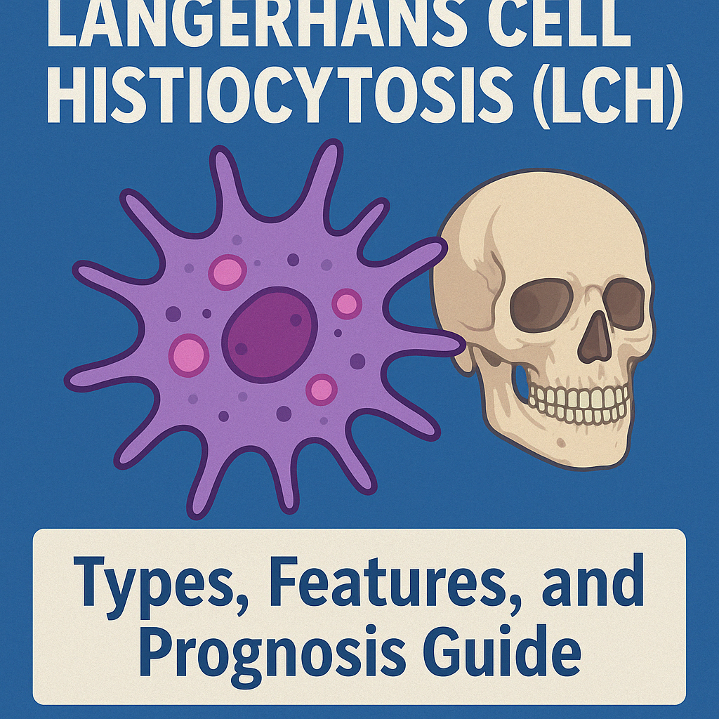
Langerhans Cell Histiocytosis (LCH) is a rare, systemic disorder characterized by clonal proliferation of abnormal Langerhans cells, affecting bone, skin, lymph nodes, and internal organs.
Orbital involvement is found in about 20–23% of LCH cases, with proptosis and superolateral bone erosion being classic findings.
🔹 Classification & History
- Previously grouped as Histiocytosis X (Eosinophilic Granuloma, Hand-Schüller-Christian, Letterer-Siwe)
- Now considered a spectrum of a single disease based on shared pathology
- Different clinical severity and prognosis depending on extent
🔹 Diagnosis
- Histology: Grooved nucleus, eosinophils, plasma cells
- EM: Birbeck granules = “tennis racket” shaped organelles
- IHC: CD1a and S100 positivity
- Imaging: CT/MRI reveals bone destruction, soft tissue mass
🔹 Clinical Manifestations & Management
- Localized lesion: biopsy, steroid injection
- Multifocal/multisystem: radiation + chemotherapy (prednisone + vinblastine)
- Most commonly affects frontal bone, causing superolateral orbital mass and displacement
🔹 Prognosis
- Worse with younger age (<2y) and multisystem involvement
- Mortality: ~55–60% under 2 years; ~15% over 3 years
- Diabetes insipidus indicates risk of chronic disease/recurrence
📋 LCH Subtypes Summary Table (English)
SubtypeKey FeaturesPrognosis
| Eosinophilic Granuloma | Localized to bone, 4–7 y/o, uni/multifocal lesions | Favorable |
| Hand-Schüller-Christian | Bone + soft tissue, proptosis + skull lesions + DI | Intermediate |
| Letterer-Siwe | Acute, systemic, multiorgan (liver, spleen, marrow) | Poor |
| Orbital LCH | Superolateral bone erosion on imaging | Depends on dissemination |
| Diagnostic Markers | CD1a(+), S100(+), Birbeck granules | Mandatory for diagnosis |
