
Tistoryview
Anatomical structure, function, distribution cells of the conjunctiva, and characteristics related to goblet cell diseases
eye_doc 2025. 4. 21. 22:55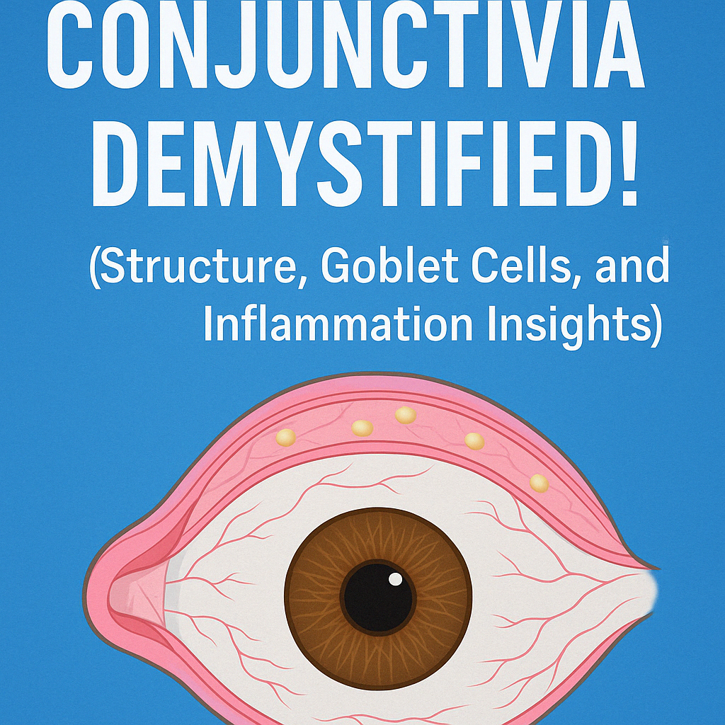
✅ Detailed Overview of the Conjunctiva – Structure, Physiology, and Clinical Significance
The conjunctiva is a transparent mucous membrane covering the anterior surface of the eye and the inner eyelid, playing a central role in tear film stabilization, ocular surface immunity, and lubrication.
🔹 1. Anatomical Subdivisions
The conjunctiva is not a homogenous layer—it is subdivided by anatomical region:
- Limbal conjunctiva: at the corneal margin, contains epithelial stem cells
- Bulbar conjunctiva: covering the globe
- Palpebral conjunctiva: lining the eyelids
- Tarsal: lies over the tarsal plate
- Orbital: associated with deeper eyelid structure
- Marginal: at the lid margin, most pain-sensitive - Fornix conjunctiva: forms the fold between bulbar and palpebral conjunctiva
🔹 2. Epithelial Features and Glycocalyx
The epithelium consists of 2–3 cell layers, increasing to 10+ near the limbus and lid margin.
Apical vesicles produce a mucin-rich glycocalyx, which ensures adherence of the tear film's mucin layer.
This is sustained by specialized crypts known as the Crypt of Henle.
🔹 3. Goblet Cells – Mucin Factories with Diagnostic Value
Goblet cells reside between epithelial cells and produce mucin for tear film stability.
- Density is highest during adolescence
- Decrease in: Vitamin A deficiency, OCP, SLK, neurotrophic ulcers
- Increase in: Atopic keratoconjunctivitis
Their loss leads to tear film instability, dry eye syndrome, and conjunctival keratinization.
🔹 4. Vascular and Neural Distribution
Conjunctival vessels receive blood from the anterior ciliary artery and form the superficial marginal plexus.
- Superficial vessels: affected in conjunctivitis (more redness distally)
- Deep vessels: congest in uveitis, scleritis (more redness near limbus)
This differential pattern helps clinicians distinguish superficial vs deep inflammation.
Sensory innervation is via the long ciliary nerve (CN V1);
- sensitivity is lower than the cornea, highest at the lid margin
🔬 Conjunctiva Overview Table (English)
| Epithelium | 2–3 layers (up to 12 near limbus/margin) | Glycocalyx for mucin adhesion / vulnerable to preservatives |
| Crypt of Henle | Goblet cell gland structure, mucin secretion | Maintains tear film / damage leads to dryness |
| Goblet Cells | 5–10% of basal cells, secrete mucin | ↓ in Vit A deficiency, OCP, SLK / ↑ in AKC |
| Vascular Pattern | Superficial plexus + deep ciliary vessels | Helps distinguish conjunctivitis vs uveitis/scleritis |
| Sensory Nerves | CN V1 (long ciliary) / deep distribution, low sensitivity | Explains pain variance (e.g. pseudomembrane removal) |